
The Equine Distal Limb
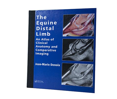
An Atlas of Clinical Anatomy and Comparative Imaging by Jean-Marie Denoix.
Watch this product and we will notify you once it is back in stock.
The Equine Distal Limb
The Equine Distal Limb – Atlas of Clinical Anatomy and Diagnostic Imaging
The Equine Distal Limb is a comprehensive atlas of the clinical anatomy of the horse's distal limbs, including the hoof, fetlock and pastern joint. It is written by Jean-Marie Denoix, a leading specialist in equine musculoskeletal anatomy and a pioneer of diagnostic ultrasonography.
About the book
This reference atlas combines anatomical dissections with detailed images from radiography, ultrasound and magnetic resonance imaging (MRI). Advanced imaging techniques require detailed anatomical knowledge; the book helps readers understand the anatomy in three dimensions.
Each section begins with an anatomical drawing by the author, followed by photographs, black-and-white diagrams and comparisons with diagnostic images.
Who is this book for?
- Veterinary surgeons and specialists investigating lameness.
- Farriers and orthopaedic specialists seeking a detailed understanding of foot and joint anatomy.
- Students and researchers in veterinary medicine and equine anatomy.
- Professionals using advanced diagnostic techniques and interpreting images.
Specifications
| Property | Details |
|---|---|
| ISBN | 978-1-84076-001-9 |
| Pages | 400 |
| Format | 29 x 24 cm |
| Language | English |
| Author | Jean-Marie Denoix |
Reference resource
With detailed images and coverage of each part of the equine distal limb, this book is a reference for professionals working with advanced lameness investigation and diagnostic imaging.